Why Choose Us?
At Edgebrook Radiology we offer :
- Quick scheduling availability to accommodate your needs.
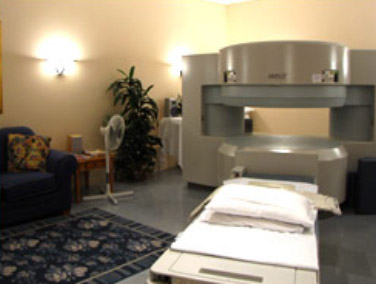
- High field and OPEN MRI to accommodate you if you are claustrophobic.
- MRI Arthrography to better visualize the inside of your joints and surrounding structures.
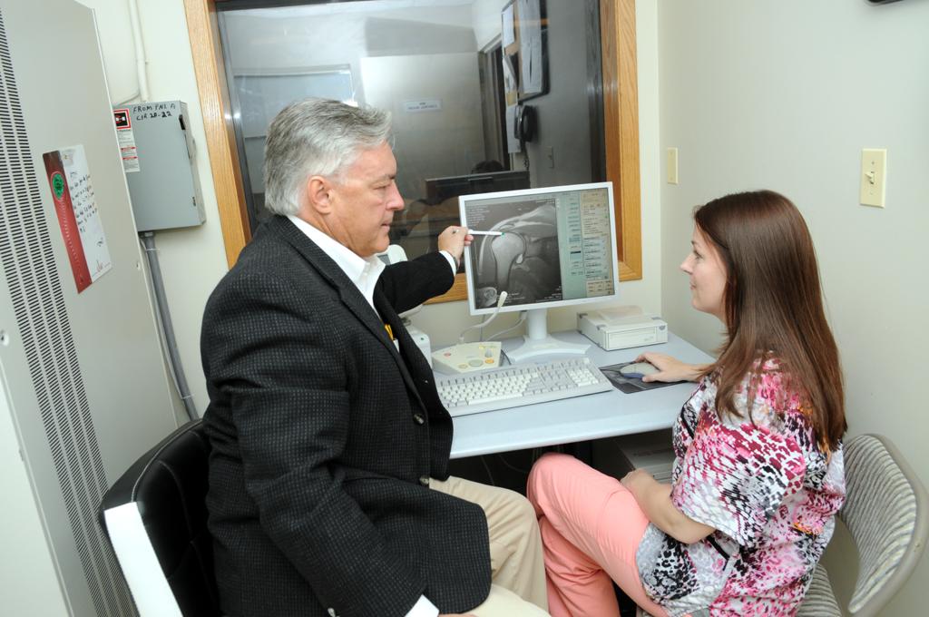
- Highly sub-specialized board-certified radiologists to monitor and interpret your exam.
- Prompt reporting of your results to your medical provider.
- Complimentary copies of your images on CD on request by your doctor.
What is MRI?
Magnetic resonance imaging, or MRI, uses a strong magnet and radio waves to provide clear and detailed diagnostic images of internal body organs and tissues. Not only does MRI avoid exposure to x-rays, but it also allows evaluation of body structures that may not be as visible with x-ray, CT, or other diagnostic imaging methods.
One special feature of MRI is its ability to obtain views of the body from any angle or direction, so that complex anatomy can be demonstrated from the most revealing perspectives. Another characteristic of MRI is its ability to characterize fine differences amongst tissues. Finally, an MRI examination can provide a broad overview of a large portion of the body, like the whole torso; or it can focus with exquisite detail on the smallest structures of, for instance, a single toe. For smaller body parts, special “imaging coils” are positioned over the area of interest to produce the most detailed pictures.
Considerable training and expertise is needed on the part of the radiologist and technologist to make the best decisions in performing the examination, and also in its correct interpretation. That is why the quality of MRI examinations obtained from different imaging centers may vary so much in their ability to provide you with the correct diagnosis. That is where the special expertise of Progressive Radiology shines. We have earned our reputation for excellence by performing specially tailored examinations for your exact clinical problem, and providing rapid, accurate interpretations.
What are some common uses of MRI?
Neuroradiology: Brain, Spine, Head and Neck
- Evaluation for stroke, tumors, multiple sclerosis, headache, vascular malformations, congenital abnormalities, bleeds, dementia
- Specialized examinations of the nerves involved in hearing, balance, vision, facial control or pain
- Studies of the pituitary gland, orbits, inner ear, salivary glands.
- Identification of aneurysms, vascular malformations, blood clots.
- Studies of the cervical, thoracic, or lumbar spine for intervertebral disk degeneration or herniation; nerve root or spinal cord compression; stenosis; tumors; abnormalities of the spinal cord; arthritis or fractures
- Soft tissue tumors in the neck or throat, abnormal lymph nodes.
- Surgical planning and follow up
Musculoskeletal: Joints, Bones and Soft Tissues
- Evaluation for arthritis, causes of pain in any joint or loss of function
- Assessment of cartilage, ligaments, tendons, and muscles
- Examination of bones for fractures, bone marrow disease, or cancer
- Characterization of abnormal fluid collections, soft tissue masses, tumors, infections of the soft tissues or bones
- Planning or follow up for surgery
- Detailed examination of the inside of joints by injection of contrast agent directly into the joint (an “arthrogram”, described in the Musculoskeletal page of our web site).
Body Imaging: Internal Organs
- Evaluation for disorders or tumors in the abdomen including the liver, adrenal glands, kidneys, pancreas, spleen, gallbladder and bile ducts
- Assessment for disorders or tumors in the pelvis including the uterus and ovaries (endometriosis, adenomyosis, fibroids, tumors, cancer, cysts, uterine anomalies); urinary bladder; pelvic floor for incontinence; urethral diverticulum; prostate cancer.
Angiography: Arteries and Veins
- Studies of the arteries and veins in the head and neck for blockage, stenosis, aneurysm, malformations, clot.
- Evaluation for aneurysm, enlargement, or narrowing of the thoracic aorta and its branches.
- Assessment of the renal arteries for stenosis as a cause of hypertension.
- Evaluation of the abdominal aorta for aneurysm.
- Arterial blood supply to the legs.
How should I prepare for an MRI?
The magnetic field is very powerful, with the ability to dislodge or warm metal objects in the body; or to disrupt the function of electronic devices. Metal objects in the body can also distort the MRI images. However, not every implanted medical device or metal object in the body will prevent you from having an MRI. It is a shame when patients assume they are unable to have an MRI because of metal in their body, when in fact often they would be able to have the study. For your safety, we have a screening process that begins when you first contact our scheduling specialist, and continues upon your arrival with the technologist and radiologist before you are put in the magnetic field. Sometimes an x-ray will be taken first to assess for the location or size of any metal foreign bodies or medical devices in your body.
Scheduling your exam
When you schedule your MRI, be sure to tell our booking specialist.
- If your body contains any metal fragments or implanted medical devices that might affect your suitability for MRI, such as a pacemaker or other electronic devices, aneurysm clip, cochlear implant, shrapnel or bullet fragment; possible metal particles in the eyes; prosthetic joints or surgical hardware
- If you might be pregnant,
- If you are claustrophobic or think you might feel overly anxious and confined in the MRI unit. You may want to ask your doctor to prescribe a sedative for use during the examination.
- If you are unable to lie still because of pain. You may want take pain medication prior to the examination.
- If your prescription mentions the use of contrast (gadolinium) or an arthrogram.
- If you have had others studies of the area we will be scanning.
Before you arrive
- For some patients who will be receiving an injection of contrast in a vein, blood test results (BUN and Creatinine) will need to be obtained.
- For some patients with implanted medical devices, wallet cards or other documents verifying their MRI compatibility will need to be obtained.
- Bring with you, or take any sedatives or pain medications so that they will be working at the time of the exam. If you do take a sedative, please be sure to have transportation arrangements so that you will not have to drive afterwards.
- If you are having an examination of the abdomen or pelvis, don’t eat for four hours prior to the examination (although medications may be taken with a small amount of water).
- Bring any relevant prior reports or images from radiology studies performed at other offices.
- Plan to arrive 30 minutes early to allow time to complete any required paperwork and review your history with the intake personnel and technologist.
- Dress in clothing that’s easy to remove, without metal, and leave your jewelry at home.
What should I expect during the exam?
On Arrival
- You will complete any necessary paperwork; review your history and safety screening questions with the technologist.
- Before your MRI exam, you will need to remove all accessories including hairpins, jewelry, eyeglasses, hearing aids, wigs, and dentures. During the exam, these metal objects may be pulled into the MRI unit, or interfere with the magnetic field, affecting the quality of the MRI images.
- Often you will be asked to change into a gown, and you will be provided with a secure area to store your belongings.
- The technologist may tape a marker on your skin to identify the location of your symptoms.
The MRI Exam
Depending on how many images are needed, the exam generally takes 15 to 45 minutes. Very detailed studies may take longer.
- You will lie down on a sliding table and be comfortably positioned.
- Sometimes a special “imaging coil” will be positioned to focus on a small anatomic area.
- Even though the technologist must leave the room, you will be able to communicate at any time using an intercom.
- If necessary, many MRI centers allow a friend or family member to stay in the room with you during the exam. They must be screened for safety in the magnetic field, and leave credit cards and electronic devices out of the examination room.
- You will be asked remain completely still during the actual imaging process. However, between pulse sequences, slight movement is allowed. Some pulse sequences require you to hold your breath briefly.
- MRI is painless.
- You will hear loud tapping or thumping during the exam. Earplugs or earphones with music will be provided.
- You may feel warmth in the area being examined. This is normal.
- Some examinations employ injection of contrast material into a vein.
After the Exam
- You may be provided with a copy of the images obtained on a CD-ROM or film.
- If your doctor has access to our network, he will be able to see the results shortly after the study is completed.
- A signed, transcribed report of our radiologist’s interpretation is provided to your doctor usually within 24 hours.