Why Choose Us?
At Edgebrook Radiology we offer:
- Same or next day availability
- Nationally registered and certified technologists with many years of ultrasound experience.
- Ultrasound technologist with additional training in performing highly specialized pediatric examinations.
- On-site board-certified radiologist to monitor and interpret exams.
- Prompt reporting of results to your medical provider.
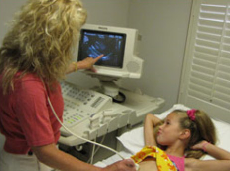
What is Ultrasound Imaging?
Ultrasound imaging (also called sonography), is uses high frequency sound waves to obtain images of the human body. The sound waves are transmitted, and then received back through a hand-held device called a transducer. A computer converts this information into images, which the skilled sonographer can observe continuously while moving the transducer over your skin, constantly changing the point of view and adjusting technical factors to demonstrate the anatomy to best advantage. A unique feature of sonography is that is involves no exposure to x-rays, and the pictures can be seen in real time. That means the patient can collaborate with the sonographer during the examination to identify tender areas or nodules that both can feel, so that the study becomes similar to a physical examination with extended vision beneath the surface of the skin. Several types of transducers are selected to provide views the wider coverage or depth, or to focus in greater detail on smaller areas closer to the skin. In general, sound waves do not pass at all through bone or air (as in gas in the gastrointestinal tract), do pass very well through water (as in a distended bladder); and provide only more limited views of deep structures or those beneath a thick layer of fat. One special transducer is the endovaginal probe, inserted into the vagina to permit a much closer view of the uterus, ovaries, cervix and young fetus.
Doppler ultrasound allows demonstration of blood flow, characterization of blood flow speed and direction, turbulent flow and periodic motion. These features are essential in examination of arteries for demonstrating adequate flow, patterns of flow that imply pathology of the neighboring soft tissues such as inflammation, inadequate blood supply, or the presence of tumors; and evaluation of veins for appropriate direction of flow and absence of blood clots. Doppler techniques are often used as an adjunct to soft tissue examinations or may be the basis of a vascular study.
What are some common uses of Ultrasound?
- Evaluation of pregnancy and fetal development.
- Examining many of the body’s internal organs, including the gallbladder or kidneys for stones; obstruction to the bile ducts or urinary tract; measurement of the liver, spleen or kidneys for size, diffuse or focal abnormalities; search for tumors; evidence of inflammation or infection.
How should I prepare for an Ultrasound?
- Wear comfortable, loose-fitting clothing.
- Examinations of the abdomen or of the abdominal aorta require that you have nothing to eat or drink for 6-8 hours prior to the exam. That includes avoidance of tea, coffee, soda, juice, or flavored waters. That is partly because the gallbladder, a common cause of right upper abdominal pain contracts when you eat, so that gallstones may be not be seen, and the gallbladder itself cannot be assessed for inflammation. Eating and drinking also introduces gas into the stomach and intestines, which blocks the view of the pancreas, aorta, and sometimes other organs. A small amount of water is allowed to take medications.
- Examinations of the kidneys, bladder, pelvis, uterus, ovaries, and for pregnancy require distension of the bladder. Drink 32 oz of water 30-45 minutes before the exam, and refrain from urinating.
What should I expect during this exam?
The examination usually takes about 30 minutes. You may change into a gown or simply loosen or remove some clothing, with care being taken to respect privacy by draping with sheets or towels so that only the necessary part of the body is exposed. Be prepared to show the technologist any lump or mass that is being investigated as part of the examination. After being positioned on the exam table, a clear gel is applied in the area being examined. This helps the transducer make contact with the skin. The gel is water-soluble, and it wipes off or can be rinsed off easily.
The technologist firmly presses the transducer against the skin and moves it back and forth to image the area of interest. Most ultrasound exams are painless. Examinations for pregnancies or of the uterus and ovaries often make use of the transvaginal transducer. Under some circumstances, a chaperone will be available to attend your exam. Sometimes the exam will be interrupted so that you can empty your bladder; or so that you can drink more water so that it is adequately full.
Often the sonographer will have the patient change position to get a better view, or to demonstrate mobility of a stone in the gallbladder or urinary bladder; hold his breath to immobilize the organs, or perform other maneuvers to change the direction of venous blood flow. Generally, the technologist is able to review the ultrasound images in real-time or, when the examination is complete and the gel is wiped off, you may be asked to dress and wait while the ultrasound images are prepared for presentation to the radiologist and the examination double-checked for quality.
Our sonographers are exceptionally well-trained and competent in conducting examinations on their own and in documenting the relevant images, which they then present to the interpreting radiologist for discussion and reporting. However, it is not uncommon for the radiologist to repeat portions of the examinations personally for more detailed evaluation of questionable findings, and to maintain the ongoing process of technologist training and quality control.